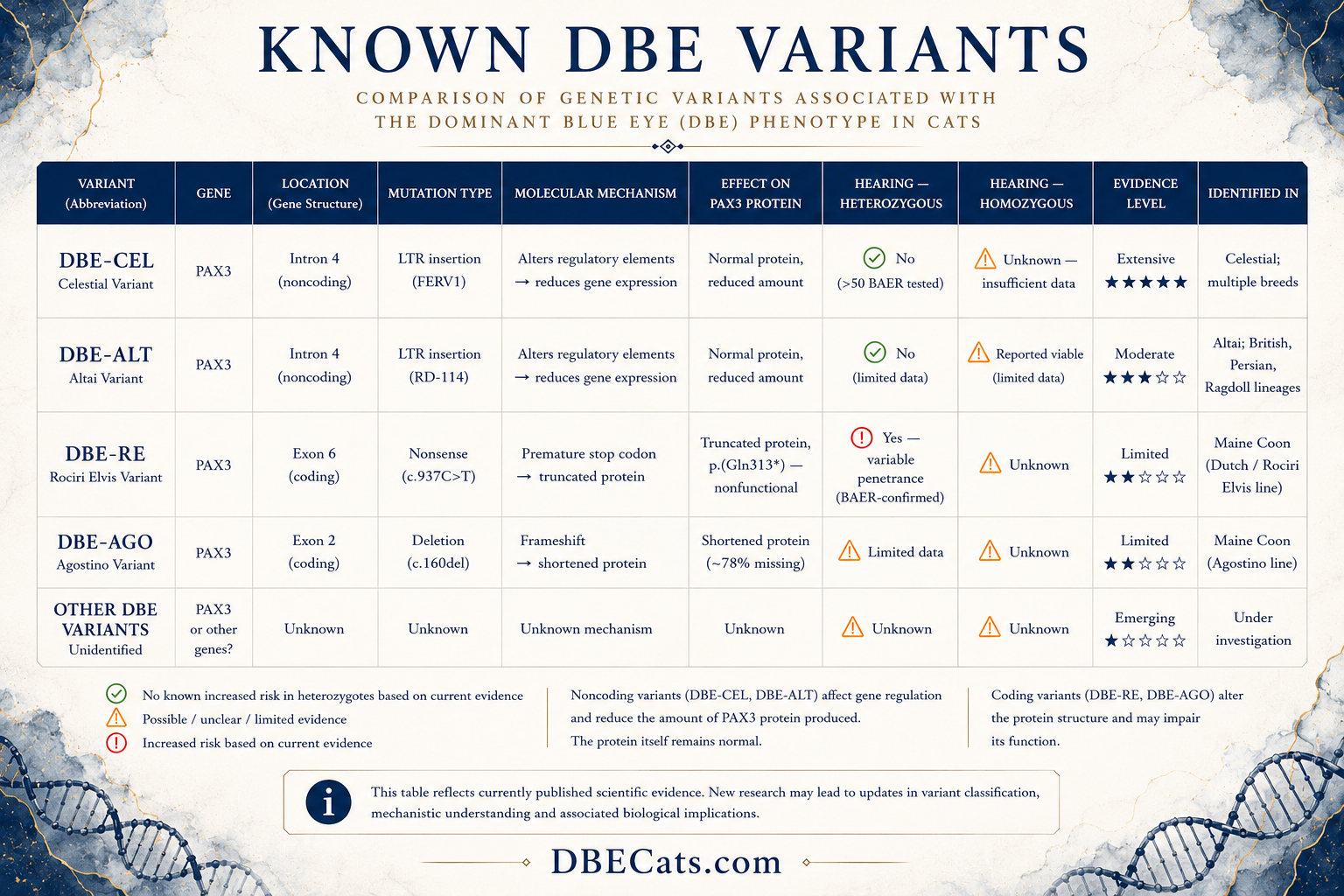
DBE Variants
Understanding the genetic diversity behind the Dominant Blue Eye phenotype.
Not all DBE variants are biologically equivalent.
1. Introduction
Understanding DBE Starts with Understanding the PAX3 Gene
Dominant Blue Eyes (DBE) is not caused by a single genetic variant. Several different genetic variants can produce a similar blue-eye phenotype, but they do not affect the PAX3 gene in the same way.¹ ² ³ ⁴
Before comparing these variants, it is important to understand how a gene is organised, what the PAX3 protein does, and why the location of a mutation can influence its biological effect.
2. What is the PAX3 Gene?
Most nucleated cells in a cat's body contain DNA organised into 38 chromosomes, arranged in 19 pairs. Chromosomes contain many genes.
The PAX3 gene contains the instructions for producing the PAX3 protein. PAX3 is a transcription factor that helps regulate the development and migration of melanocytes — the pigment-producing cells of the skin, coat, eyes and inner ear — in part by acting as an upstream regulator of the MITF gene.¹ ³
The PAX3 gene, associated with several Dominant Blue Eye (DBE) variants, is located on chromosome C1.¹
3. How Is a Gene Structured?
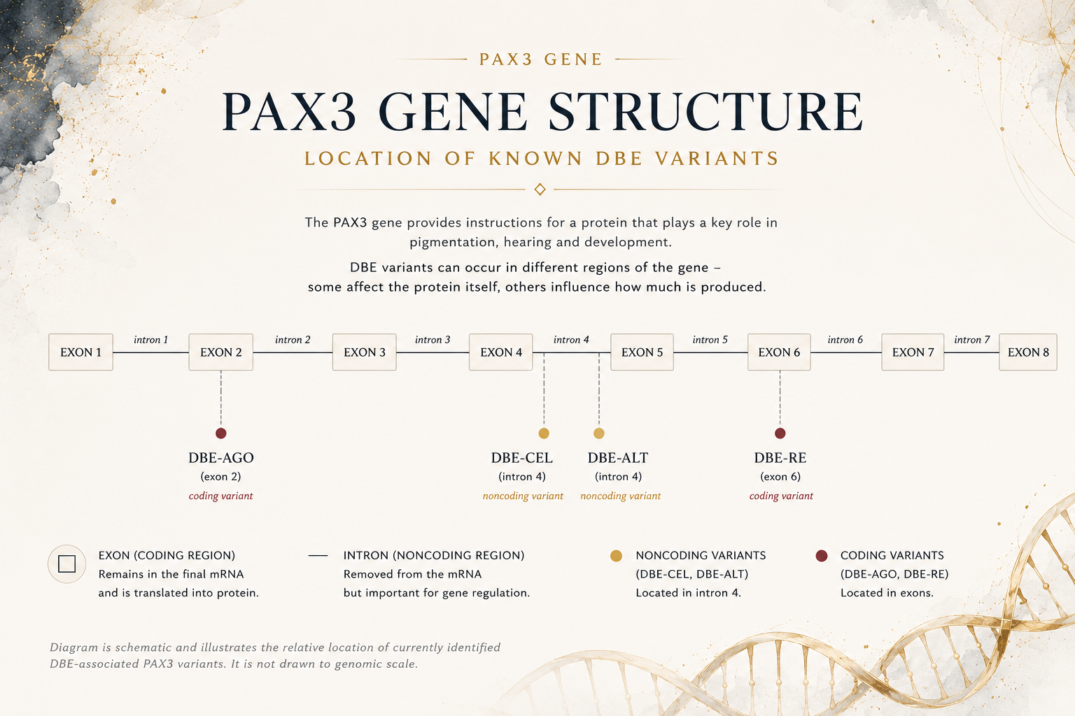
A gene is a continuous region of DNA, but protein-coding genes are usually divided into two types of segments: exons and introns.
Exons
Exons contain the sequence used to build the protein. These regions remain in the mature messenger RNA (mRNA) and their coding portion is translated into the final protein.
Introns
Introns are located between exons and are removed from the initial RNA transcript during splicing. Although introns are not translated into protein, some intronic regions contain regulatory elements that may influence gene expression or RNA processing.
Because of these different functions, mutations in exons and introns can have very different biological consequences.
4. Why does the mutation location matter?
The location of a mutation is just as important as the mutation itself. Variants occurring in different regions of the same gene can influence gene function through entirely different molecular mechanisms.
5. Understanding the Difference
Non-coding variants
Non-coding variants occur outside the protein-coding sequence, including within introns. They do not directly alter the protein-coding DNA sequence, but they may affect gene regulation or RNA processing. DBE-CEL and DBE-ALT are distinct LTR insertions within intron 4 of PAX3, in a region containing conserved regulatory elements. Their precise effects on PAX3 expression and protein abundance have not yet been demonstrated experimentally.¹
Coding variants
Coding variants occur within exons, the regions that encode the protein. Because they directly alter the sequence that is translated, they may change the protein’s amino acid sequence, reduce its function, or prevent it from functioning normally. The DBE-RE and DBE-AGO variants are examples of protein-changing variants in PAX3.³ ⁴
6. Why This Matters
Grouping all DBE variants together can lead to oversimplified conclusions. Identifying the specific variant provides a more accurate framework for interpreting scientific literature, assessing potential biological effects, and making informed breeding decisions.
References
Abitbol M, Couronné A, Dufaure de Citres C, Gache V. A PAX3 insertion in the Celestial breed and certain feline breeding lines with dominant blue eyes. Animal Genetics. 2024;55(4):670–675. doi:10.1111/age.13433
Abitbol M, Dufaure de Citres C, Rudd Garces G, Lühken G, Lyons LA, Gache V. Different Founding Effects Underlie Dominant Blue Eyes (DBE) in the Domestic Cat. Animals. 2024;14(13):1845. doi:10.3390/ani14131845
Rudd Garces G, Farke D, Schmidt MJ, Letko A, Schirl K, Abitbol M, Leeb T, Lyons LA, Lühken G. PAX3 haploinsufficiency in Maine Coon cats with dominant blue eyes and hearing loss resembling the human Waardenburg syndrome. G3 Genes|Genomes|Genetics. 2024;14(9):jkae131. doi:10.1093/g3journal/jkae131
Abitbol M, Cloquell A, Kaczmarska A, Holmes K, Lühken G, Macaulay K. Dominant blue eyes in Maine Coon cats: New PAX3 variant and updated phenotypic data. Animal Genetics. 2025;56(3):e70020. doi:10.1111/age.70020
Known PAX3 Variants
Several independent PAX3 variants have now been identified as being associated with the Dominant Blue Eyes phenotype.
Although these variants affect the same gene, they differ in their genomic location, mutation type and predicted molecular consequences. Some occur within protein-coding exons, while others are located in non-coding introns. As a result, they are not expected to affect PAX3 function in the same way.
Current evidence indicates that these variants should be interpreted individually rather than considered as a single genetic entity.
The currently characterised PAX3-associated DBE variants are presented below.
7. DBE-CEL
Overview
DBE-CEL is a non-coding intronic variant of the PAX3 gene, first described by Abitbol et al. (2024). It consists of a FERV1 LTR insertion within intron 4 of PAX3, close to conserved non-coding elements involved in the regulation of PAX3 expression. It is currently the best-characterised non-coding variant associated with the Dominant Blue Eye (DBE) phenotype.
Location
The DBE-CEL variant is located within intron 4 of the PAX3 gene on chromosome C1. Because introns are non-coding regions, the mutation does not become part of the final protein sequence.
Molecular Mechanism
DBE-CEL is a non-coding intronic variant with a proposed regulatory effect.
Because the insertion lies outside the protein-coding sequence, it is not predicted to alter the amino acid sequence of the PAX3 protein directly. Its position close to regulatory elements in intron 4 instead suggests it may influence PAX3 expression during development. This effect is inferred from the variant's location and has not yet been directly measured in DBE-CEL cats.
This distinguishes DBE-CEL from coding variants such as DBE-RE and DBE-AGO, which alter the protein-coding sequence and are predicted to produce abnormal or truncated PAX3 proteins.
Associated Phenotype
Published cases have associated DBE-CEL with a range of eye pigmentation patterns, including:
bilateral blue eyes
odd eyes
sectoral heterochromia
latent (non-expressing) cats
The degree of expression appears to be variable, suggesting that additional genetic or developmental factors may influence the final phenotype.
Hearing
BAER testing of DBE-CEL heterozygous cats has not shown evidence of an association between the variant and deafness (Abitbol, Couronné, et al., 2024).
Among 47 BAER-tested DBE-Celestial cats, three were diagnosed with unilateral deafness and none with bilateral deafness. The three unilaterally deaf cats had concurrent ear conditions unrelated to DBE and the authors concluded that the DBE-CEL variant did not appear to be associated with deafness in heterozygous cats.
At present, this distinguishes DBE-CEL from some coding PAX3 variants, which have been associated with hearing impairment.
Current Evidence
One genetically confirmed DBE-CEL/DBE-CEL kitten has been reported in the scientific literature. The kitten was genetically confirmed as homozygous for the DBE-CEL insertion; it was white, had a cleft palate and died at birth, and limb abnormalities were also reported by the breeder. What is established for this individual is its genotype, not the cause of these findings: no study has demonstrated that DBE-CEL homozygosity causes them. A single case cannot establish causation, and it remains unknown whether any, some or all of these abnormalities were related to the genotype, to other genetic or developmental factors, or to unrelated causes.
No genetically confirmed living homozygous DBE-CEL cat has been described in the scientific literature to date.
This absence of published cases should not be interpreted as evidence that living DBE-CEL homozygotes do not exist. It indicates only that no such cat has yet been genetically documented and described in the peer-reviewed literature.
Unpublished breeder reports cannot establish the phenotype or viability of DBE-CEL homozygotes unless the cats are individually genotyped and their clinical findings are documented.
References
Abitbol M. et al. (2024). A PAX3 insertion in the Celestial breed and certain feline breeding lines with dominant blue eyes.Animal Genetics, 55(4), 670–675.
8. DBE-ALT
Overview
DBE-ALT is a non-coding intronic variant of the PAX3 gene, first described by Abitbol et al. (2024). It consists of an RD-114 LTR insertion within intron 4 of PAX3, close to conserved non-coding elements involved in the regulation of PAX3 expression, and represents an independently arising Dominant Blue Eye (DBE) variant.
Although DBE-ALT is located within the same intron as DBE-CEL, it originated from a different endogenous retroviral insertion and should therefore be considered a distinct genetic variant rather than a subtype of DBE-CEL.
Location
The DBE-ALT variant is located within intron 4 of the PAX3 gene on chromosome C1.
Molecular Mechanism
DBE-ALT is a non-coding intronic variant with a proposed regulatory effect.
Because the insertion lies outside the protein-coding sequence, it is not predicted to alter the amino acid sequence of the PAX3 protein directly. Like DBE-CEL, its position close to regulatory elements in intron 4 instead suggests it may influence PAX3 expression during development. This effect is inferred from the variant's location and has not yet been directly measured in DBE-ALT cats.
Although both variants affect intron 4, they arose independently through different retroviral insertion events and should be regarded as separate molecular variants.
Associated Phenotype
Published cases have associated DBE-ALT with a range of eye pigmentation phenotypes, including:
bilateral blue eyes
odd eyes
sectoral heterochromia
latent (non-expressing) cats
As with other DBE variants, expression appears to be variable, and additional genetic or developmental modifiers are likely to influence the final phenotype.
Hearing
Based on the currently available published data, hearing loss has not been reported in heterozygous DBE-ALT cats.
Deafness has been associated with compound heterozygosity and homozygosity.
Relatively few genetically characterised animals have been described for these combined genotypes, and further studies are needed to better define the clinical spectrum of this variant.
Current State of Knowledge
DBE-ALT was first identified in 2024 as one of the independently arising PAX3 variants associated with the Dominant Blue Eye phenotype.
Unlike DBE-CEL, surviving homozygous (RD/RD) DBE-ALT cats have been reported. Three were described, all white or almost white: one (Nanotigr line) was reported deaf by its owner, while two (Altai line) were considered to hear normally by their breeder. As BAER testing was not performed on these two, unilateral deafness could not be excluded. Their existence confirms that homozygous DBE-ALT cats are viable, but the number of genetically confirmed homozygotes remains very small, and BAER-tested cases are needed to define the clinical spectrum accurately.
Current knowledge of DBE-ALT remains considerably more limited than that of DBE-CEL, and further research is needed to fully characterise this variant.
References
Abitbol M. et al. (2024). Different Founding Effects Underlie Dominant Blue Eyes (DBE) in the Domestic Cat Animals, 14(13), 1845.
9. DBE-RE
Overview
DBE-RE is a coding nonsense variant of the PAX3 gene, first described by Rudd Garces et al. (2024) in Maine Coon cats.³
Unlike DBE-CEL and DBE-ALT, which are intronic variants with proposed regulatory effects, DBE-RE occurs within the protein-coding sequence of PAX3. The variant introduces a premature stop codon and is predicted to disrupt normal PAX3 function.
Location
DBE-RE is located in exon 6 of the PAX3 gene on chromosome C1.³
How Does It Work?
DBE-RE is a coding nonsense variant, PAX3:c.937C>T, with the predicted protein-level consequence PAX3:p.(Gln313*).
The nucleotide substitution changes the codon for glutamine at amino-acid position 313 into a premature stop codon. The variant is therefore predicted to result in PAX3 haploinsufficiency.
This differs from DBE-CEL and DBE-ALT, which are intronic insertions located near regulatory elements and do not alter the PAX3 protein-coding sequence.
Associated Phenotype
Cats carrying the DBE-RE variant have been reported with:
two blue eyes;
heterochromia iridis, commonly described as odd eyes;
minimal white spotting;
sensorineural hearing loss.
Latent, non-expressing DBE-RE cats carrying the variant without displaying the blue-eye phenotype have also been reported.⁴
The phenotype partially resembles human Waardenburg syndrome, which is associated with reduced PAX3 function and includes abnormalities of pigmentation and sensorineural hearing loss.³
Hearing
DBE-RE has been associated with hearing impairment in heterozygous cats.
In the original study, all eight heterozygous DBE-RE cats examined using BAER testing had unilateral or bilateral hearing loss. A subsequent 2025 study BAER-tested six additional heterozygous DBE-RE cats, all of which had normal bilateral hearing.
The available evidence therefore supports an association between DBE-RE and an increased risk of hearing impairment, but deafness is not fully penetrant. Its prevalence cannot yet be determined from the limited number of BAER-tested animals.
Current State of Knowledge
Current knowledge of DBE-RE is based on two peer-reviewed studies involving cats from the same Maine Coon lineage.
The original study investigated eight stillborn kittens from the pedigree. Six were heterozygous for DBE-RE and two were homozygous for the wild-type allele. None was homozygous for DBE-RE, and the authors concluded that the deaths were not correlated with the PAX3 genotype.³
The subsequent 2025 study identified latent, non-expressing DBE-RE cats and heterozygous DBE-RE cats with normal bilateral hearing. These findings demonstrate variable expression of both the eye-pigmentation and hearing phenotypes within the lineage.⁴
To date, no genetically confirmed homozygous DBE-RE cat has been described in the scientific literature. Consequently, the biological effects of inheriting two copies of the DBE-RE variant remain unknown.
References
Rudd Garces G, Farke D, Schmidt MJ, Letko A, Schirl K, Abitbol M, Leeb T, Lyons LA, Lühken G. PAX3 haploinsufficiency in Maine Coon cats with dominant blue eyes and hearing loss resembling the human Waardenburg syndrome. G3: Genes|Genomes|Genetics. 2024;14(9):jkae131. doi:10.1093/g3journal/jkae131.
Abitbol M, Cloquell A, Kaczmarska A, Holmes K, Lühken G, Macaulay K. Dominant blue eyes in Maine Coon cats: New PAX3 variant and updated phenotypic data. Animal Genetics. 2025;56(3):e70020. doi:10.1111/age.70020.
10. DBE-AGO
Overview
DBE-AGO is the fourth currently characterised PAX3 variant associated with the Dominant Blue Eyes phenotype. It was first described by Abitbol et al. (2025) in the Agostino Maine Coon lineage.
Unlike DBE-CEL and DBE-ALT, which are intronic variants with proposed regulatory effects, DBE-AGO is a coding frameshift variant predicted to produce a severely truncated PAX3 protein.
Location
DBE-AGO is located in exon 2 of the PAX3 gene on chromosome C1.
How Does It Work?
DBE-AGO is caused by a single-base deletion, PAX3:c.160del.
The deletion shifts the reading frame and introduces a premature stop codon. It is predicted to produce a truncated PAX3 protein, PAX3:p.(His54ThrTer108), lacking approximately 78% of the amino acids present in the wild-type protein.
This differs from DBE-CEL and DBE-ALT, which are intronic insertions located near regulatory elements and do not alter the PAX3 protein-coding sequence.
Associated Phenotype
In the original publication, DBE-AGO was associated with:
two blue eyes;
heterochromia iridis, commonly described as odd eyes;
minimal white spotting.
Some cats also showed more extensive white spotting caused by a separate white-spotting allele of the KIT gene.
All 13 genetically characterised DBE-AGO cats in the study were heterozygous for the variant.
Hearing
No deafness was reported by breeders in the Agostino lineage.
However, none of the genetically characterised DBE-AGO cats underwent formal BAER testing. It is therefore not currently possible to determine whether DBE-AGO is associated with unilateral or bilateral hearing impairment.
The absence of breeder-reported deafness should not be interpreted as equivalent to a normal BAER result.
Current State of Knowledge
DBE-AGO is currently described in a single peer-reviewed study.
All 13 genetically characterised DBE cats from the Agostino lineage were heterozygous for PAX3:c.160del. The variant was absent from the non-DBE relatives examined and from all control cat genomes (n > 300).
No latent, non-expressing DBE-AGO cat was identified in the studied family, although the number of animals examined remains limited.
No genetically confirmed homozygous DBE-AGO cat has been described in the scientific literature. Its association with hearing impairment, full phenotypic spectrum and biological consequences in homozygous cats therefore remain unknown.
References
Abitbol M, Cloquell A, Kaczmarska A, Holmes K, Lühken G, Macaulay K. Dominant blue eyes in Maine Coon cats: New PAX3 variant and updated phenotypic data. Animal Genetics. 2025;56(3):e70020. doi:10.1111/age.70020.
11. DBE-CDC
Overview
DBE-CDC (also listed as DBE5 in some commercial testing panels) is a variant currently offered for genetic testing by at least one laboratory but not yet described in the peer-reviewed scientific literature. As of 2026, no published study, preprint or OMIA entry characterises this variant.
Unlike DBE-CEL, DBE-ALT, DBE-RE and DBE-AGO — each of which has been formally described and peer-reviewed — DBE-CDC has not been independently validated in the scientific literature. Its designation, following the naming convention used for other DBE variants, most likely refers to the breeding line in which it was first identified rather than to a published genomic characterisation.
Location and Molecular Mechanism
Not established in the peer-reviewed literature.
Because no scientific publication describing DBE-CDC is currently available, its genomic location, mutation type and molecular mechanism cannot be stated. It is not known whether it is a coding or non-coding variant, nor how it may affect the PAX3 protein or its expression.
Associated Phenotype and Hearing
No peer-reviewed phenotypic or BAER hearing data have been published for DBE-CDC. Any association with eye colour, white spotting or hearing status therefore cannot currently be assessed against the scientific literature.
How to Interpret a DBE-CDC Result
A laboratory result reporting DBE-CDC (or DBE5) confirms that the laboratory has detected a specific genetic marker in the tested cat. However, because the variant has not been peer-reviewed, a positive result cannot be interpreted in the same evidence-based framework as the four published variants.
The absence of a publication does not prove that the variant does not exist genetically; it means only that its biological effect, phenotype and health implications have not yet been scientifically documented. Until such data are published, DBE-CDC results should be treated with appropriate caution, and no claims regarding health, hearing or breeding safety should be based on them.
12. DBE-XHW
Overview
DBE-XHW (also listed as DBE6 in some commercial testing panels) is a variant currently offered for genetic testing by at least one laboratory but not yet described in the peer-reviewed scientific literature. As of 2026, no published study, preprint or OMIA entry characterises this variant.
It has been associated with Chinese domestic cats / domestic shorthairs. Unlike DBE-CEL, DBE-ALT, DBE-RE and DBE-AGO — each of which has been formally described and peer-reviewed — DBE-XHW has not been independently validated in the scientific literature. Following the naming convention used for other DBE variants, its designation most likely refers to the line or population in which it was first identified rather than to a published genomic characterisation.
Location and Molecular Mechanism
Not established in the peer-reviewed literature.
Because no scientific publication describing DBE-XHW is currently available, its genomic location, mutation type and molecular mechanism cannot be stated. It is not known whether it is a coding or non-coding variant, nor how it may affect the PAX3 protein or its expression.
Associated Phenotype and Hearing
No peer-reviewed phenotypic or BAER hearing data have been published for DBE-XHW. Any association with eye colour, white spotting or hearing status therefore cannot currently be assessed against the scientific literature.
How to Interpret a DBE-XHW Result
A laboratory result reporting DBE-XHW (or DBE6) confirms that the laboratory has detected a specific genetic marker in the tested cat. However, because the variant has not been peer-reviewed, a positive result cannot be interpreted in the same evidence-based framework as the four published variants.
The absence of a publication does not prove that the variant does not exist genetically; it means only that its biological effect, phenotype and health implications have not yet been scientifically documented. Until such data are published, DBE-XHW results should be treated with appropriate caution, and no claims regarding health, hearing or breeding safety should be based on them.